

Aortic stenosis is a heart valve condition in which the aortic valve becomes narrowed, making it harder for blood to leave the heart and reach the rest of the body. As the narrowing becomes more significant, the heart may need to work harder to maintain adequate blood flow.
For patients searching for aortic stenosis treatment in Bangalore, understanding the severity of the valve disease is an important first step. Treatment is not determined by symptoms alone. Doctors consider the valve’s structure, the degree of narrowing, heart function, age, overall health, symptoms, and other medical conditions before recommending monitoring, medication, transcatheter treatment, or surgery.
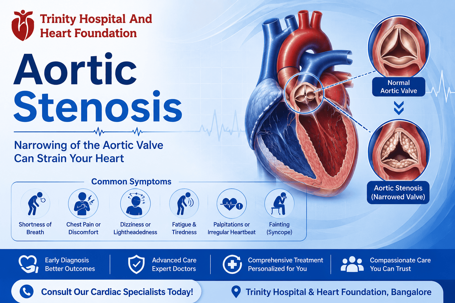
The condition may progress gradually, and some people have few symptoms during the early stages. When symptoms develop, they may include breathlessness, chest discomfort, dizziness, fainting, fatigue, or reduced ability to perform usual activities. Severe symptomatic aortic stenosis generally requires evaluation for valve replacement. Current ACC/AHA guidance recommends that patients with severe valve disease being considered for intervention receive multidisciplinary assessment and that the choice between surgical and transcatheter replacement should involve individualized, shared decision-making.
At Trinity Hospital and Heart Foundation, Basavanagudi, Bengaluru, patients can access cardiac diagnostic, interventional, and cardiothoracic services, including echocardiography, coronary assessment, TAVI/TAVR, balloon valvuloplasty, and surgical valve replacement pathways.
The aortic valve sits between the left ventricle and the aorta. It opens when the heart pumps so blood can move from the heart into the body’s main artery.
In aortic stenosis, the valve opening becomes narrowed. This is sometimes described as aortic narrowing, although the medically precise term refers to narrowing of the aortic valve rather than the aorta itself.
When the valve becomes significantly narrowed:
NHLBI explains that aortic stenosis restricts blood flow through the aortic valve and makes the heart work harder.
The aortic stenosis causes can vary depending on age and the underlying valve abnormality.
Common causes include:
Calcium deposits can accumulate on the aortic valve over time, causing the valve leaflets to become stiff and narrow.
Some people are born with a bicuspid aortic valve, meaning the valve has two leaflets rather than the usual three. This can increase the likelihood of developing aortic valve narrowing.
Previous rheumatic fever can damage heart valves and may contribute to valve narrowing.
Some people are born with structural abnormalities affecting the aortic valve.
Infections and other forms of valve damage can also affect valve function.
The Trinity Hospital TAVR page identifies age-related calcification, bicuspid aortic valve, rheumatic disease or valve infection, and progressive valve narrowing among factors associated with aortic stenosis.
Aortic Stenosis Signs and Symptoms
Early aortic stenosis may cause few noticeable symptoms. As narrowing becomes more significant, symptoms may become easier to notice during physical activity.
Common aortic stenosis signs and symptoms include:
These symptoms are not specific to valve disease and can occur with other forms of cardiac disease. Therefore, symptoms alone cannot confirm aortic stenosis.
Trinity Hospital advises cardiac evaluation for persistent breathlessness, chest discomfort, dizziness, fainting, or unexplained fatigue.
Severe untreated valve disease can eventually contribute to heart failure because the heart has to work harder to pump blood through the narrowed valve.
Possible progression may involve:
Aortic valve narrowing → increased workload on the heart → changes in heart muscle → reduced cardiac function → symptoms of heart failure
This does not mean that every person with aortic stenosis develops congestive heart failure. The risk depends on the severity and duration of the disease and other individual factors.
Symptoms associated with advanced heart failure may include:
The symptoms of cardiac heart failure should be evaluated medically because they can have several possible causes.
A detailed cardiac assessment is necessary to determine whether valve narrowing is mild, moderate, or severe.
Evaluation may include:
The doctor reviews symptoms, medical history, risk factors and physical findings such as a heart murmur.
ECG
An electrocardiogram records the heart’s electrical activity and can provide supporting information about the heart.
Echocardiography
An echocardiogram uses ultrasound to assess:
Echocardiography is central to assessing aortic stenosis severity. Trinity Hospital lists 2D echocardiography among its cardiac diagnostic services.
Depending on the clinical situation, doctors may consider CT imaging, stress testing, cardiac catheterization, or other investigations.
ACC/AHA guidance recommends correlating history and examination findings with non-invasive testing and using additional non-invasive or invasive testing when the initial findings do not provide enough clarity.
Treatment decisions depend heavily on disease severity.
Doctors assess factors such as:
Severe aortic stenosis with symptoms or reduced ventricular systolic function may require valve replacement. In selected patients, earlier intervention can be considered based on factors such as very severe stenosis, rapid progression, exercise-test findings, or biomarkers.
This is why an echocardiogram report should be interpreted by a qualified cardiologist rather than based on one measurement in isolation.
The appropriate aortic stenosis treatment depends on severity, symptoms, valve anatomy, age, surgical risk, heart function, and other health conditions.
People with mild or moderate disease and no indication for valve intervention may be monitored through:
The frequency of follow-up depends on the individual’s condition.
There is currently no medicine that physically opens a severely narrowed aortic valve.
However, medications may be used to manage:
Heart failure medications may be appropriate when a patient has heart failure or another clinical indication, but they do not reverse the mechanical narrowing of the aortic valve.
NHLBI notes that medicines may help manage symptoms or other conditions associated with valve disease, while valve repair or replacement may eventually be required in appropriate patients.
Transcatheter Aortic Valve Replacement (TAVR), also called TAVI, is a minimally invasive valve replacement procedure.
A replacement valve is delivered through a catheter and positioned within the diseased aortic valve.
NHLBI describes TAVR as a catheter-based procedure that replaces a diseased or narrowed aortic valve without traditional open-heart surgery.
Trinity Hospital currently lists Transcatheter Aortic Valve Implantation (TAVI) as an interventional cardiology service. Its dedicated TAVR page states that the procedure is considered for suitable patients based on valve anatomy, age, health conditions, and other clinical factors.
TAVR may be considered depending on:
The decision should be individualized rather than based solely on age.
Surgical aortic valve replacement involves replacing the diseased valve through cardiac surgery.
It may be appropriate when:
Trinity Hospital’s cardiology department lists Valve Replacement (MVR / AVR) and valve repair surgery under cardiothoracic surgery.
The choice between surgical valve replacement and TAVR requires careful evaluation by the appropriate cardiac team.
Balloon valvuloplasty uses a catheter with a balloon to widen a narrowed aortic valve.
It may be used in selected clinical circumstances, but it is not generally considered a permanent replacement for a severely diseased valve.
Trinity Hospital lists balloon valvuloplasty among its catheter-based cardiac procedures.
| Factor | TAVR/TAVI | Surgical Valve Replacement |
| Approach | Catheter-based | Cardiac surgery |
| Chest opening | Does not require traditional open-heart surgery | Usually requires surgical access |
| Recovery | Often shorter than traditional surgery, depending on patient | Usually longer |
| Suitability | Depends on anatomy, age, risk and clinical factors | Depends on anatomy, age, risk and other surgical needs |
| Other cardiac surgery | Limited compared with surgery | Can be combined with other cardiac procedures when required |
| Decision | Individualized | Individualized |
Neither approach is automatically suitable for every patient. Current guidelines recommend shared decision-making and multidisciplinary evaluation for severe valve disease being considered for intervention.
What Happens If Aortic Stenosis Is Not Treated?
The progression of aortic stenosis varies from person to person.
Significant untreated disease can eventually contribute to:
NHLBI notes that untreated heart valve disease can lead to heart failure and other serious complications.
This is why regular follow-up is important even when symptoms are mild or absent.
Aortic Stenosis and Related Terms: What Do They Mean?
Some patients encounter different medical terms while researching their diagnosis.
Narrowing of the aortic valve that restricts blood flow from the heart.
Aortic valve stenosis ICD-10 terminology is used for medical classification and documentation. The specific code depends on the clinical diagnosis and coding system being used. It should not be used by patients as a substitute for a medical diagnosis.
The phrase aortic stenosis ICD 10 is commonly searched when patients or healthcare professionals are looking for the appropriate diagnostic classification. Coding should be confirmed against the patient’s documented diagnosis and the applicable coding guidelines.
Supravalvular aortic stenosis is a different and less common form of obstruction located above the aortic valve, rather than narrowing of the valve itself.
Aortoiliac stenosis refers to narrowing in the aorta or iliac arteries, usually in the vascular system, and is not the same condition as aortic valve stenosis.
These distinctions matter because treatment depends on the exact location and cause of the narrowing.
Trinity Hospital and Heart Foundation in Basavanagudi provides an integrated cardiac care pathway involving diagnostics, interventional cardiology and cardiothoracic surgery.
The hospital’s current cardiology services include:
The department describes a team-based approach involving cardiology consultants, nursing staff, diagnostic teams and referring doctors, with second opinions and external referrals when required.
Dr. B. G. Muralidhara is listed by Trinity Hospital as a Senior Interventional Cardiologist and Chief Cardiologist.
His profile states that he has more than three decades of clinical experience, with a career in cardiology beginning in 1994. His qualifications include MBBS, MD in General Medicine, DM in Cardiology and FICC. The profile also reports experience involving more than 10,000 angioplasties, 60,000 coronary angiograms and 2,000 pacemaker implantations.
His listed areas of expertise include:
For patients with suspected valve disease, the hospital’s TAVR page states that Dr. Muralidhara evaluates patients for TAVR suitability based on valve anatomy, overall health and individual clinical factors.
Consider a cardiac evaluation if you have:
Symptoms such as severe or sudden chest pain, significant breathlessness, fainting, or other acute cardiac symptoms require prompt medical assessment.
The cost of aortic stenosis treatment in Bangalore varies according to the patient’s condition and treatment pathway.
Factors may include:
A specific treatment cost should only be discussed after clinical evaluation because the appropriate treatment and investigations vary between patients.
Aortic stenosis can range from mild disease requiring monitoring to severe valve narrowing requiring valve replacement. The appropriate treatment depends on symptoms, valve severity, heart function, anatomy, age, surgical risk and other individual factors.
For patients seeking aortic stenosis treatment in Bangalore, a detailed cardiac evaluation can help determine whether continued monitoring, medical management, TAVR/TAVI, surgical valve replacement, or another approach is appropriate.
Trinity Hospital and Heart Foundation in Basavanagudi provides diagnostic, interventional and cardiac surgical pathways for patients requiring evaluation of heart valve disease.
 24 Hour Emergency Aid